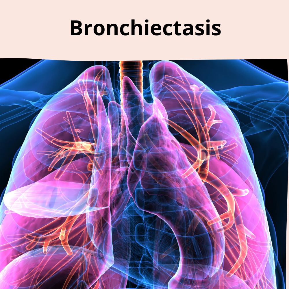
Bronchiectasis is a chronic condition affecting the airways of the lungs. The airways (bronchi) walls get damaged, become scarred and thickened, and ultimately lose the ability to clear mucus. As a result of this, the bronchi get permanently enlarged, which leads to serious health problems.
Types of Bronchiectasis
- Cylindrical or tubular bronchiectasis – The most common form shows a smooth uniform enlargement of the bronchi with loss of normal tapering of the airways.
- Varicose bronchiectasis – The dilated bronchi are interspersed with sites of relative narrowing, giving rise to a beaded appearance.
- Cystic or saccular bronchiectasis – is the most severe form that is often found in patients with cystic fibrosis and is characterized by clusters of cysts in the dilated bronchi.
- Traction bronchiectasis – It results from mechanical traction of the fibrotic lung surrounding the bronchi and is seen in conditions like interstitial lung disease.
Causes:
- Damage to the walls of the bronchi is the main etiological factor of bronchiectasis. It could happen due to a lung infection or other health conditions.
- Bronchiectasis is usually the primary lung manifestation of the following recurring conditions that affect the airways:
- Severe pneumonia
- Pertussis (whooping cough)
- Viral infections (due to measles, adenovirus, influenza virus)
- Tuberculosis
- Allergic bronchopulmonary aspergillosis
- Mucociliary dysfunction and autoimmune disorders can also lead to bronchiectasis.
- Airway blockage due to growth or noncancerous tumors, traction, and cystic fibrosis can also lead to bronchiectasis.
- It also occurs as a secondary complication to other lung diseases such as :
- Chronic obstructive pulmonary disease (COPD)
- Emphysema
- Bronchitis
- Bronchiolitis
- Interstitial lung disease
- Chronic pulmonary aspiration
Risk factors of Bronchiectasis
- Genetic disorders – Cystic fibrosis, primary ciliary dyskinesia, Kartagener’s syndrome, Young’s syndrome, and Tracheobronchomegaly increase the risk of bronchiectasis.
- Impaired host defenses – Weakened or absent immune system responses due to primary immunodeficiency, HIV/AIDS, Job’s syndrome, or prolonged use of immunosuppressive drugs also raises the risk.
- Autoimmune disorders – Rheumatoid arthritis, Sjogren syndrome, ulcerative colitis, and Crohn’s disease increases the rate of bronchiectasis.
- Gender – Women are more susceptible than men due to increased inflammatory responses during menstrual cycles and pregnancy. In children, there is a stark contrast, as boys are at a higher risk than girls.
- Age – People above 65 years of age are more susceptible as the risk generally increases with age.
- Other health conditions – Allergic bronchopulmonary aspergillosis, chronic pulmonary aspiration, and hiatal hernia put the person at a higher risk.
- Environmental factors – Inhalation of ammonia and other toxic gases, heavy alcohol use, drug abuse, and pollution aggravates other lung infections and increases the risk of bronchiectasis.
Signs and symptoms of Bronchiectasis
- Chronic cough
- Coughing up varying quantities of sputum containing yellow or green mucus for months or years
- Coughing up blood in the absence of sputum
- Shortness of breath
- Wheezing
- Chest pain
- Fatigue
- Recurring chest infection
- Chronic sinus inflammation
- Nail clubbing (rare)
Complications:
Severe bronchiectasis leads to health complications such as:
- Respiratory failureAtelectasis
- Heart failure
Diagnosis: How is Bronchiectasis diagnosed?
- The doctor usually examines the patient’s family history to look for signs of genetic disorders implicated in bronchiectasis.
- Bronchiectasis is generally suspected if the patient has been suffering from regular coughing that produces sputum.
- Diagnostic tests –
- Chest CT scan – A chest computed tomography (CT) scan provides images of the airways and shows the extent and location of the lung damage.
- Chest x-ray – Abnormal regions in the lungs and thickened, irregular airway walls are detected via this test.
- Blood tests – Complete blood count is done to check the presence of infections and determine the levels of infection-fighting blood cells. It also helps to identify any underlying condition that could lead to bronchiectasis.
- Sputum cultures – The sputum coughed up by the patient is tested in a laboratory to check for the presence of bacteria or fungi that can cause lung infections.
- Lung function tests – Spirometry and walking tests are done to assess and monitor the functioning impairment of the lungs.
- Sweat test – The salt content in the patient’s sweat is tested as high salt levels can be caused by cystic fibrosis. Genetic testing is generally recommended if the results are positive.
- Bronchoscopy – Chest Physicians can detect blockages in the airways by inserting a flexible tube into the airways via the nose or the mouth.
Treatment:
- Early diagnosis and treatment of bronchiectasis are essential as this prevents exacerbation of the disease.
- Treating the underlying condition responsible for bronchiectasis and preventing complications is the primary rationale behind the treatment. Removal of the excess mucus from the lungs is also an important goal of the treatment as this provides favorable conditions for bacterial growth.
- Medications –
- Antibiotics – Azithromycin, Tobramycin, etc., are given to treat recurrent bacterial infections and exacerbations.
- Expectorants and mucus-thinning medicines – Guaifenesin, acetylcysteine, etc. help to loosen and cough up the mucus.
- Bronchodilators – albuterol, levalbuterol, etc. are used to relax the muscles around the airways and make it easier to breathe.
- Inhaled corticosteroids – Beclometasone dipropionate therapy reduces sputum production and decreases airway constriction, especially if the patient has wheezing.
- Chest Physical Therapy (CPT) – An electric chest clapper is used to pound the chest back and over to loosen the mucus that the patient can then cough up.
- Hydration – Drinking plenty of water keeps the airway mucus moist and slippery, making it easier to cough up.
- Oxygen therapy – To raise low blood levels.
- Surgery – If medicines remain ineffective, the damaged part of the lung or airway is surgically removed.
Prevention:
- Living with it –
- Exercising regularly
- Quitting smoking
- Staying hydrated
- Eating a healthy diet
- Preventing it –
- Having a flu vaccine every year
- Getting pneumococcal vaccine
- Avoiding toxic fumes and gases
When to see a doctor? :
It is advised to see a pulmonologist if persistent or recurrent cough and sputum production is purulent and large volume. It costs around Rs. 500 to 2K to consult with a pulmonologist in India.
References:
